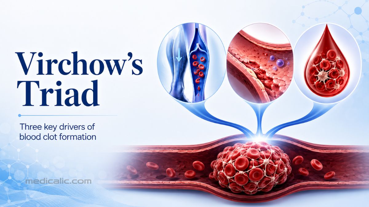
A blood clot usually does not form because of one isolated event. More often, several changes inside the body come together and create the right environment for clotting.
Doctors explain this process through Virchow’s triad, a medical framework built around three factors:
- Slow or abnormal blood flow
- Injury or dysfunction of a blood vessel’s inner lining
- An increased tendency of the blood to clot
The concept is especially useful for understanding deep vein thrombosis (DVT) and pulmonary embolism (PE). Together, these conditions are known as venous thromboembolism, or VTE.
Identifying these risk factors early may create opportunities for prevention and timely clinical assessment. However, Virchow’s triad is not a test and cannot tell you whether you currently have a clot.
What Is Virchow’s Triad?
Virchow’s triad describes three biological conditions that promote thrombosis, the formation of a blood clot inside a blood vessel. [1]
| Component | What it means | Common examples |
|---|---|---|
| Venous stasis | Blood moves too slowly or pools in a vein | Bed rest, limited mobility, long travel, paralysis |
| Endothelial injury or dysfunction | The blood vessel lining becomes damaged or starts promoting clotting | Surgery, trauma, inflammation, catheters, smoking |
| Hypercoagulability | The blood develops an increased tendency to clot | Cancer, pregnancy, estrogen therapy, inherited clotting disorders |
A simple way to remember the triad is:
Not every person has all three factors to the same degree. One strong factor may be enough to create substantial risk, especially when it overlaps with smaller or temporary risks. The Centers for Disease Control and Prevention notes that the likelihood of DVT or PE rises further when multiple risk factors occur together. [2]
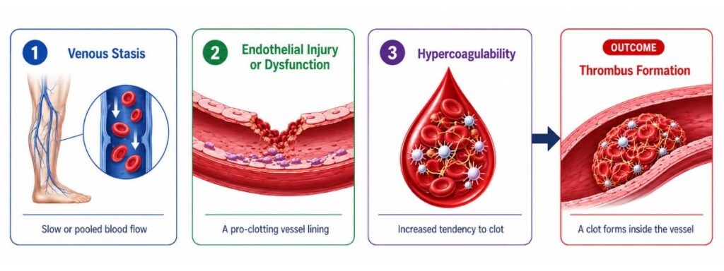
1. Venous Stasis: When Blood Flow Slows Down
Blood is meant to keep moving.
In the legs, muscle contractions squeeze the veins and help move blood back toward the heart. One-way valves inside the veins keep it from flowing backward.
When you remain still for a long time, this pumping action becomes less effective. Blood may begin to pool, particularly in the deep veins of the legs. [5]
Slow flow makes clotting more likely because activated clotting proteins are not carried away or diluted as efficiently. Blood cells and coagulation factors also remain near the vessel wall for longer.
Common causes of venous stasis include:
- Prolonged bed rest
- Hospitalization
- Recovery after major surgery
- Paralysis or limited mobility
- A leg cast
- Long-distance travel
- Heart failure
- Pressure on pelvic veins during pregnancy
- A previous DVT that damaged the venous valves
Sitting for several hours does not automatically mean that you will develop a clot. The risk becomes more important when immobility is combined with factors such as recent surgery, active cancer, pregnancy, estrogen use, obesity, older age, or a previous VTE.
Why DVT Often Starts Near Venous Valves
Many DVTs begin in small pockets around the valves of deep leg veins. Blood flow is naturally slower in these areas.
When flow becomes extremely sluggish, local oxygen levels may fall and the endothelium may become activated. This can attract inflammatory cells and clotting proteins, creating a favorable environment for a fibrin-rich clot to grow.
2. Endothelial Injury or Dysfunction
The endothelium is the thin layer of cells lining the inside of every blood vessel.
It is not simply a passive wall. A healthy endothelium actively helps:
- Maintain smooth blood flow
- Prevent unnecessary platelet activation
- Control inflammation
- Support the body’s natural anticoagulant systems
- Regulate fibrinolysis, the process that breaks down clots
When the endothelium is physically damaged or becomes dysfunctional, it may shift from an anticoagulant surface to a pro-clotting one.
It can begin releasing or activating substances that encourage platelets, inflammatory cells, and clotting proteins to gather at the affected area.
Causes of Direct Vessel Injury
Physical damage may occur because of:
- Major surgery
- Trauma
- Bone fractures
- Severe burns
- Central venous catheters
- Other vascular devices
Dysfunction Without a Visible Injury
A vessel does not need to be cut or torn for its lining to become prothrombotic. Endothelial dysfunction may also occur with:
- Severe infection or sepsis
- Chronic inflammation
- Low oxygen levels
- Diabetes
- Cancer
- Chronic vascular disease
- Cigarette smoking
Smoking primarily affects the endothelial component of Virchow’s triad by promoting oxidative stress, inflammation, and impaired vascular function. Its effects may also extend into hypercoagulability by increasing platelet activity and shifting the blood toward a more pro-clotting state.
3. Hypercoagulability: When Blood Clots Too Easily
Your body normally maintains a careful balance between forming clots, limiting their growth, and breaking them down.
Hypercoagulability means that this balance has shifted toward thrombosis.
Some causes are inherited. Others develop because of illness, medication, hormonal changes, or major physical stress.
Inherited Causes
Common inherited thrombophilias include: [3]
- Factor V Leiden: Makes factor V resistant to inactivation by activated protein C
- Prothrombin G20210A mutation: May increase the amount of prothrombin in the blood
- Antithrombin deficiency: Weakens an important natural anticoagulant pathway
- Protein C deficiency
- Protein S deficiency
Acquired Causes
More common acquired causes include:
- Active cancer
- Antiphospholipid syndrome
- Pregnancy and the postpartum period
- Estrogen-containing birth control
- Hormone replacement therapy
- Major surgery or trauma
- Severe infection or inflammation
- Heparin-induced thrombocytopenia
- Certain myeloproliferative blood disorders
Not everyone who develops a clot needs thrombophilia testing. Testing is considered selectively when the result is likely to change management, such as decisions about anticoagulation, future prevention, pregnancy or hormone use, or family counselling.
It is generally not recommended after a VTE clearly provoked by major surgery. Because anticoagulant treatment and the timing of testing can affect some results, a clinician should decide whether and when testing is appropriate.
How the Three Parts of Virchow’s Triad Work Together
Virchow’s triad becomes most useful when you stop viewing its components as three separate lists and look at how they interact.
Major Surgery
Surgery can activate every part of the triad:
- Stasis: Bed rest and reduced movement slow venous circulation.
- Endothelial injury: Surgery causes tissue and blood vessel trauma.
- Hypercoagulability: The body temporarily increases clotting activity in response to injury and inflammation.
This is why hospitals assess VTE risk and may recommend early walking, intermittent compression devices, compression stockings, or anticoagulant medication for selected patients.
Cancer
Cancer may increase clotting risk through several mechanisms:
- Tumors may release procoagulant substances.
- Cancer can trigger systemic inflammation.
- A mass may compress a vein and slow blood flow.
- Illness may reduce mobility.
- Surgery and some cancer treatments may increase risk.
- Central venous catheters may irritate the vessel wall.
Because cancer can affect all three parts of the triad, people receiving cancer treatment may have a substantially increased risk of VTE. [4]
Pregnancy and the Postpartum Period
Pregnancy is another clear example:
- The enlarging uterus may slow venous return from the legs.
- Pregnancy naturally increases several clotting factors.
- Delivery (particularly cesarean delivery) may cause tissue and vascular injury.
These changes help protect against excessive bleeding during childbirth, but they also temporarily increase thrombosis risk. The CDC notes that pregnancy-associated risk continues during the period after delivery. [6]
Severe Infection
Serious infection may activate the endothelium, increase inflammation, stimulate coagulation, and disrupt normal blood flow.
Researchers sometimes describe this interaction between immunity and clotting as immunothrombosis. It helps the immune system contain infection, but excessive activation may contribute to harmful clot formation.
Virchow’s Triad, DVT, and Pulmonary Embolism
Deep vein thrombosis (DVT) usually forms in a deep vein of the leg or pelvis. If part of the clot breaks away and lodges in a pulmonary artery, it causes a pulmonary embolism (PE), which can obstruct blood flow through the lungs, strain the heart, and become life-threatening.
Does Virchow’s Triad Cause Symptoms?
Virchow’s triad itself does not cause symptoms. It describes the conditions that make clotting more likely.
Symptoms appear only if a clot forms and begins interfering with blood flow.
Possible DVT Symptoms
Symptoms usually affect one leg or arm and may include:
- Swelling
- Pain or tenderness
- Warm skin
- Redness or discoloration
Some DVTs produce mild symptoms or no noticeable symptoms at all.
Possible Pulmonary Embolism Symptoms
A PE may cause:
- Sudden, unexplained shortness of breath
- Chest pain that becomes worse with a deep breath or cough
- A rapid or irregular heartbeat
- Coughing up blood
- Lightheadedness
- Fainting
- Very low blood pressure
Sudden breathing difficulty, chest pain, coughing up blood, or fainting requires emergency medical attention. In the United States, call 911.
Can Virchow’s Triad Diagnose a Blood Clot?
No. Virchow’s triad explains why thrombosis may occur, but it cannot confirm or rule out DVT or PE.
Because several conditions can cause similar symptoms, clinicians first estimate the probability of VTE using the medical history, physical examination, and often a validated clinical assessment tool such as the Wells score. [7]
Depending on the clinical probability, evaluation may include:
- A D-dimer blood test
- Duplex or compression ultrasound for suspected DVT
- CT pulmonary angiography for suspected PE
- A ventilation-perfusion scan in selected patients
A D-dimer test cannot confirm VTE by itself. When the clinical probability is low or VTE is considered unlikely, a negative result can help rule it out. A positive result is not diagnostic and generally requires further evaluation with imaging.
Compression ultrasound is commonly used for suspected DVT, while CT pulmonary angiography is commonly used for suspected PE. A ventilation-perfusion scan may be used when CT pulmonary angiography is unsuitable.
How Understanding Virchow’s Triad May Help With Prevention
You cannot control every risk factor, but you can take practical steps to reduce avoidable risks.
Keep Blood Moving
Follow your medical team’s instructions about getting out of bed and walking after surgery or hospitalization.
During long journeys, change position regularly and move your feet and ankles. People with additional risk factors should ask a clinician whether they need specific travel precautions.
Discuss Your Personal Risk
Tell your healthcare team if you have:
- A previous DVT or PE
- A strong family history of blood clots
- Active cancer
- A recent operation or major injury
- Pregnancy or recent childbirth
- Estrogen-containing medication
- Long-term immobility
- A known clotting disorder
Follow Prescribed Prevention
Compression devices and anticoagulant medicines can reduce risk in appropriate patients, but they are not suitable for everyone.
Use them only as prescribed and report unusual bleeding, black stools, vomiting blood, severe headaches, or other concerning symptoms while taking an anticoagulant.
Do Not Self-Treat a Suspected Clot
Do not start aspirin or a blood thinner on your own because you think you may have DVT or PE.
These medicines can cause serious bleeding, and aspirin is not an appropriate substitute for professional evaluation or prescribed anticoagulation. Seek medical care instead.
Frequently Asked Questions
They are venous stasis, endothelial injury or dysfunction, and hypercoagulability.
It explains the biological conditions that promote thrombosis, particularly DVT and pulmonary embolism.
A common mnemonic is SHE:
1. Stasis
2. Hypercoagulability
3. Endothelial injury
Not necessarily. One dominant factor may combine with smaller or temporary risks to promote thrombosis.
The principles may also contribute to arterial thrombosis, but arterial clots are more commonly associated with atherosclerotic plaque disruption, platelet activation, and high-shear blood flow. [8]
The Bottom Line
Virchow’s triad explains how three interacting mechanisms can promote thrombosis. It helps organize blood-clot risk but cannot diagnose DVT or pulmonary embolism. Possible DVT symptoms require prompt medical assessment, while possible pulmonary embolism symptoms require emergency care.
Medical References
Update History
Medically reviewed by Dr. Rahul Kushwaha.
Originally written by Dr. Jubair Islam.
Your answer helps us improve the clarity and usefulness of our health content.