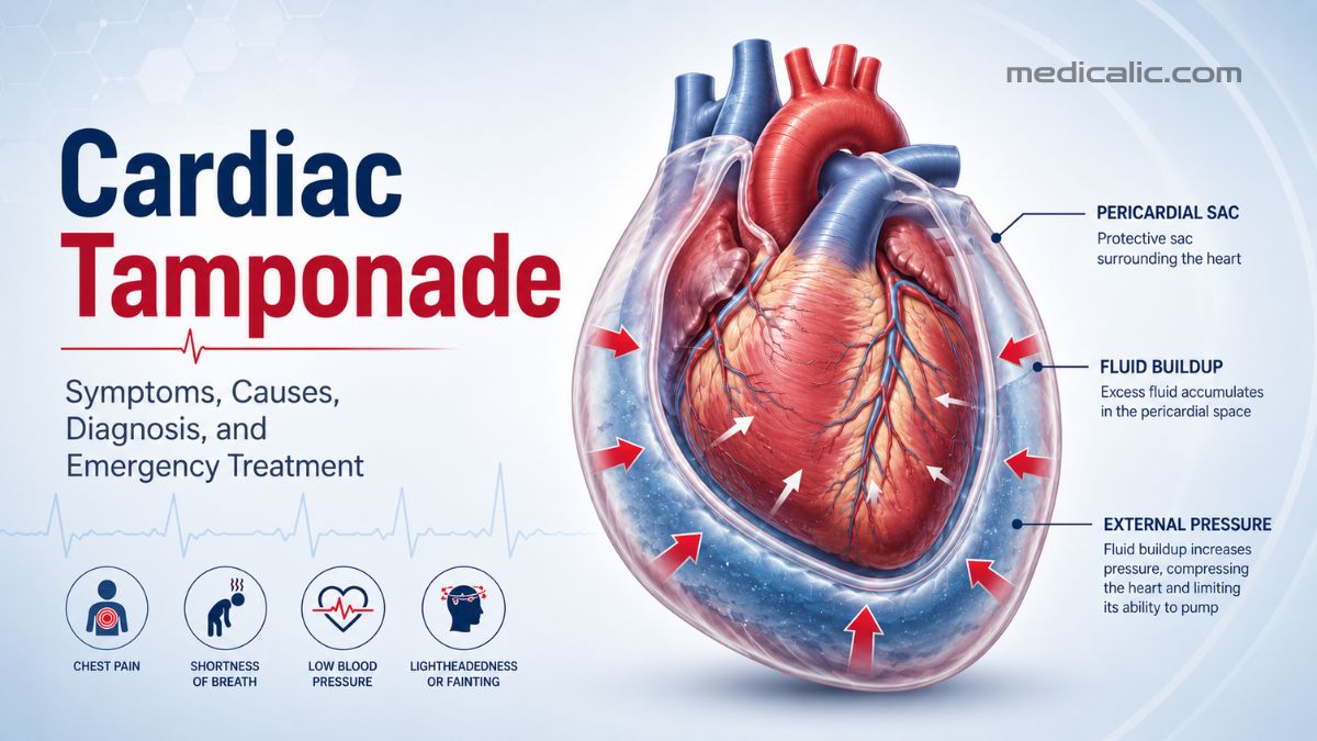
The heart needs enough space to relax and refill between beats, not just contract and pump blood forward. Cardiac tamponade occurs when fluid or blood collects inside the sac surrounding the heart and creates enough pressure to restrict that filling.[1] As a result, less blood reaches the brain and other organs.
Tamponade is more than simply “fluid around the heart.” What defines the condition is the effect of that fluid on cardiac filling and circulation. Without prompt treatment, it can quickly become life-threatening.
What Is Cardiac Tamponade?
The heart is enclosed by a thin, two-layered sac called the pericardium. A small amount of fluid normally sits between these layers, allowing them to move smoothly as the heart beats.
Problems arise when additional fluid, blood, pus, or other material collects within this confined space. As pressure rises, the chambers have less room to expand and fill.
With less blood entering the heart, less is pumped out with each beat. Blood pressure can fall, the heart rate often increases, and the organs may no longer receive enough oxygen-rich blood.
The speed of accumulation can be more important than the total volume. As little as 200–300 mL may cause tamponade if it collects rapidly, whereas a slowly developing effusion may reach 1–2 liters before heart filling becomes severely impaired.[2]
Cardiac Tamponade vs. Pericardial Effusion
A pericardial effusion is an abnormal collection of fluid around the heart. Some effusions are small, stable, and discovered incidentally during imaging without affecting circulation.
Tamponade develops only when pressure within the pericardial space begins to interfere with cardiac filling:
- Pericardial effusion: Excess fluid is present around the heart.
- Cardiac tamponade: Pressure from the collection is restricting heart filling and blood flow.
For this reason, treatment decisions depend on symptoms, circulation, and evidence of cardiac compression rather than fluid size alone.
Cardiac Tamponade Symptoms and Warning Signs
Symptoms can begin suddenly or develop gradually over hours, days, or longer. They may include:
- Shortness of breath or rapid breathing
- Chest pressure, discomfort, or fullness
- A fast or pounding heartbeat
- Dizziness, weakness, or fainting
- Difficulty breathing while lying flat
- Anxiety, restlessness, confusion, or drowsiness
As circulation deteriorates, blood pressure may fall, the pulse may weaken, and urine output may decrease. The skin can become pale, cool, or mottled, and severe cases may lead to fainting or loss of consciousness.
When severe, cardiac tamponade can cause obstructive shock because external pressure prevents the heart from filling adequately.
What Is Beck’s Triad?
The classic bedside pattern associated with cardiac tamponade is Beck’s triad:
- Low blood pressure
- Distended neck veins
- Muffled or distant heart sounds
The complete triad is often absent, so doctors do not wait for all three findings before evaluating a suspected case.[3]
Another possible finding is pulsus paradoxus, defined as a fall in systolic blood pressure of more than 10 mmHg during inspiration.[4] Although it can strengthen clinical suspicion, pulsus paradoxus is not specific to cardiac tamponade and does not appear in every patient.
What Causes Cardiac Tamponade?
Tamponade can result from disease, injury, bleeding, or complications of medical treatment. Causes include:
- Blunt or penetrating chest trauma
- Bleeding after heart surgery
- Complications of catheter ablation, cardiac catheterization, pacemaker placement, or other cardiac procedures
- Cancer involving the pericardium
- Viral, bacterial, tuberculous, or inflammatory pericarditis
- Advanced kidney failure and uremia
- Autoimmune diseases such as lupus
- Rupture of the heart wall after a heart attack
- Aortic dissection leaking into the pericardial space
- Bleeding around the heart that is worsened by anticoagulants, particularly after trauma or a cardiac procedure
The most common causes vary by region and clinical setting. Procedure-related bleeding and malignancy account for many cases in specialist centers, while tuberculosis remains an important consideration in areas where the infection is more prevalent.
How Is Cardiac Tamponade Diagnosed?
Cardiac tamponade is a clinical diagnosis, meaning no single symptom, examination finding, or imaging result is enough on its own.[8] Doctors assess the person’s symptoms, blood pressure, heart rate, circulation, medical history, physical findings, and imaging together.
An echocardiogram, or ultrasound of the heart, is usually the first imaging test. It can detect fluid and show whether pressure is affecting the heart’s chambers or normal blood flow.
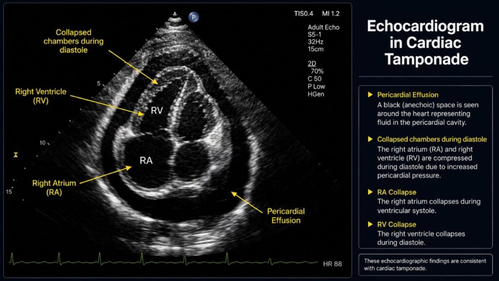
Findings that support tamponade can include:[5]
- Right-atrial collapse during ventricular systole
- Right-ventricular collapse during diastole
- A dilated inferior vena cava with limited respiratory variation
- Marked breathing-related changes in blood flow through the heart
The size of the collection alone does not establish the diagnosis. A large, slowly developing effusion may be tolerated, while a smaller or localized collection—particularly after surgery—can cause severe circulatory compromise.
Depending on the situation, doctors may also use:
- Electrocardiogram: Sinus tachycardia, low-voltage QRS complexes, or electrical alternans may appear. None of these findings confirms or excludes tamponade by itself.
- Chest X-ray: A large, gradually developing effusion may produce an enlarged cardiac silhouette, while the X-ray can remain normal in acute cases.
- CT, cardiac MRI, or transesophageal echocardiography: These tests can help when a standard echocardiogram is inconclusive or the collection is localized.
When the patient is unstable, further testing must not delay emergency treatment.
How Is Cardiac Tamponade Treated?
Treatment requires urgent relief of pressure within the pericardial space. Depending on the cause and clinical circumstances, drainage is performed through image-guided pericardiocentesis or surgery.
For many patients, pericardiocentesis is the preferred approach. A clinician inserts a needle and catheter into the pericardial space to remove fluid.[6] Draining even a modest amount can quickly improve cardiac filling, blood pressure, and breathing.
The catheter may remain in place temporarily to continue drainage and monitor for reaccumulation. Collected fluid can also be analyzed for infection, cancer cells, inflammation, or blood.
When pericardiocentesis is appropriate, echocardiographic or fluoroscopic guidance improves needle placement and procedural safety.
Surgery may be the safer option when tamponade results from:
- Traumatic bleeding
- Clotted blood
- Purulent infection
- Rupture of the heart or aorta
- A difficult or localized collection
- Repeated fluid accumulation
A surgeon may create a pericardial window, allowing fluid to drain continuously into another body cavity.
Carefully selected intravenous fluids and medications can provide temporary circulatory support while urgent drainage is arranged. These measures do not relieve the compression and must never delay definitive treatment.
Positive-pressure ventilation can reduce venous return and worsen hemodynamics before drainage. It is therefore avoided when possible in an unstable patient.[7] Diuretics also require caution because lowering circulating volume can further impair cardiac filling.
The underlying cause must be treated as well. Depending on the diagnosis, this may involve antibiotics, tuberculosis therapy, dialysis, cancer treatment, anti-inflammatory medication, or surgical repair.
Recovery and Outlook
Many patients feel noticeably better once the pressure around the heart has been relieved. Long-term outcomes, however, depend largely on the underlying cause, the severity of circulatory collapse, and how quickly treatment began.
Follow-up can involve repeat echocardiograms, drain monitoring, fluid analysis, and ongoing treatment of the condition responsible for the effusion. Recurrence is more likely when the cause is cancer, infection, or persistent inflammation.
Cardiac tamponade cannot always be prevented. Close monitoring of a known pericardial effusion can help identify worsening pressure before severe circulatory compromise develops.
After heart surgery or another cardiac procedure, new shortness of breath, chest discomfort, weakness, or fainting should be reported promptly.
When to Call Emergency Services
Call 911 in the United States or the appropriate emergency number in your country if someone:
- Has severe difficulty breathing
- Faints or becomes less responsive
- Develops confusion or unusual drowsiness
- Has cold or clammy skin
- Experiences chest symptoms with marked weakness, dizziness, or a rapid heartbeat
Symptoms beginning after chest trauma, heart surgery, catheter ablation, pacemaker placement, or another cardiac procedure require especially urgent attention.
Medical References
Update History
Medically reviewed by Dr. Rahul Kushwaha.
Originally written by Medicalic.
Your answer helps us improve the clarity and usefulness of our health content.