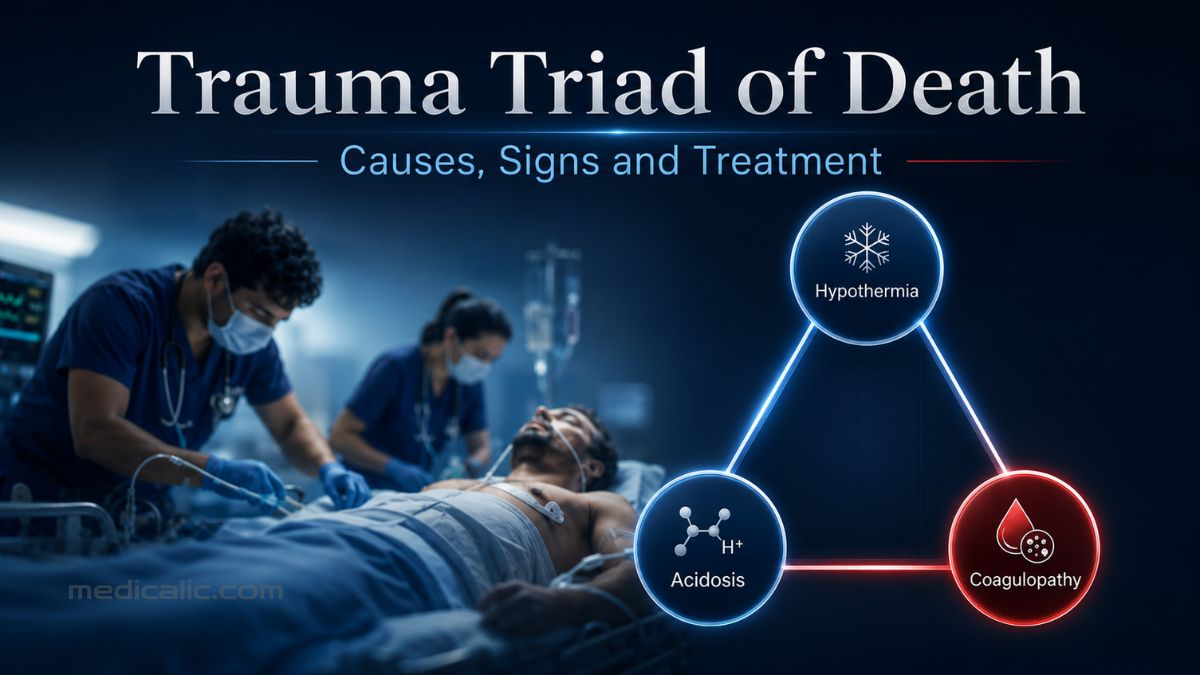
After a severe injury, the visible wound may be only part of the emergency. Inside the body, blood loss can set off a chain reaction: tissues lose oxygen, body temperature falls, and the blood becomes less able to clot. Each problem strengthens the next, allowing bleeding and shock to worsen with alarming speed.
This process is called the trauma triad of death. It involves three interconnected conditions:
- Hypothermia: dangerously low body temperature
- Metabolic acidosis: increasing acidity caused largely by inadequate tissue perfusion
- Coagulopathy: impaired formation or maintenance of effective blood clots
The term does not mean that death is inevitable. It identifies a rapidly worsening emergency that may still be interrupted with prompt hemorrhage control and coordinated trauma care.
What Is the Trauma Triad of Death?
The trauma triad of death, also called the lethal triad, describes a self-reinforcing physiological crisis in severely injured patients.
As bleeding continues, circulating blood volume, oxygen delivery, and body heat fall. At the same time, severe tissue injury and shock can trigger trauma-induced coagulopathy before substantial dilution occurs. Poor tissue perfusion promotes metabolic acidosis, while hypothermia, acidosis, ongoing blood loss, dilution, and low ionized calcium can further impair hemostasis and worsen hemorrhage.[1]
The result is a vicious circle:
No single test confirms the trauma triad. Trauma teams identify it from the combined pattern of hemorrhage, shock, falling temperature, and impaired coagulation. Major trauma guidelines emphasize early detection because traumatic coagulopathy may already be present when the patient reaches the hospital.
How the Cycle Develops
The components do not follow a fixed sequence.
Major bleeding commonly drives the process. As circulating blood volume falls, less oxygen reaches the tissues. Lactate often rises, metabolic acidosis develops, and organs begin to function less effectively.
At the same time, the patient may lose heat through blood loss, wet clothing, cold surroundings, exposure during examination or surgery, anesthesia, and the administration of unwarmed fluids or blood products.
Coagulopathy may also begin immediately because of the injury and shock themselves, not simply because the patient later becomes cold or receives large amounts of intravenous fluid. Trauma can disrupt platelets, fibrinogen, clot formation, clot breakdown, and the lining of blood vessels. Hypothermia, acidosis, dilution, low calcium, and continued hemorrhage then intensify that disruption.
So, the key clinical lesson is not to wait for a textbook sequence. The three problems may emerge almost simultaneously.
The Three Components of the Trauma Triad
Hypothermia
In severely injured trauma patients, hypothermia generally refers to a core body temperature below 35°C or 95°F. However, even a smaller temperature drop can matter when the patient is already bleeding and unstable.[2]
Normal coagulation depends on temperature-sensitive chemical reactions and properly functioning platelets. As the body becomes colder, those reactions slow and platelet function deteriorates. Hypothermia can therefore increase blood loss while making resuscitation more difficult.
Trauma teams try to prevent this early by removing wet clothing, limiting unnecessary exposure, warming the room, using forced-air warming blankets, and warming intravenous fluids and blood products. Current European guidance recommends rapid heat-loss prevention and active warming toward normal body temperature.
Metabolic Acidosis
In major trauma, metabolic acidosis usually reflects inadequate tissue perfusion rather than an isolated acid-base disturbance.
When circulation is inadequate, cells cannot produce energy normally. Lactate and other markers of tissue hypoperfusion rise, while impaired liver and kidney blood flow reduces the body’s ability to correct the disturbance.
Clinicians assess the problem through blood pH, lactate, bicarbonate, and base deficit, which some laboratories report using the related base-excess value. More importantly, they follow how these measurements change during resuscitation.
Acidosis also weakens coagulation enzyme activity. But correcting the laboratory pH alone does not solve the underlying emergency. Treatment must restore perfusion by stopping the bleeding, supporting oxygen delivery, and improving circulation and ventilation.
Trauma-Induced Coagulopathy
In the early bleeding phase, coagulopathy means the patient cannot form or maintain an effective clot.
This process is more complex than a simple shortage of clotting factors. Severe injury and shock can alter:
- Platelet activity
- Thrombin production
- Fibrinogen concentration and function
- Fibrin clot strength
- Fibrinolysis, or clot breakdown
- Endothelial function within blood vessels
Later, ongoing blood loss, low temperature, acidosis, low calcium, and excessive crystalloid fluid can make clotting even less effective.
During massive hemorrhage, red blood cell transfusion supports oxygen-carrying capacity but does not by itself replace depleted or dysfunctional platelets, fibrinogen, and other coagulation components. Depending on bleeding severity and test results, the patient may also require plasma, platelets, fibrinogen-containing products, calcium, or other targeted therapy.
Trauma-induced coagulopathy is not identical in every patient. Excessive clot breakdown may dominate in some patients, while others may develop suppressed fibrinolysis or a more prothrombotic state later in the course of injury.
How Trauma Teams Recognize the Triad
A patient does not need to have dramatically abnormal results in all three areas before treatment begins. Trauma teams repeatedly evaluate the injury pattern, active or suspected bleeding, vital signs, mental status, temperature, laboratory results, and response to resuscitation.
Concerning findings may include:
- Uncontrolled external bleeding or suspected internal hemorrhage
- Falling blood pressure or persistent circulatory instability
- Rapid pulse or breathing
- Confusion, agitation, unusual drowsiness, or loss of consciousness
- Pale, cold, or clammy skin
- Falling core temperature
- Increasing lactate or worsening base deficit
- Low blood pH
- Prolonged PT or INR
- Low fibrinogen or platelet count
- Low ionized calcium
Where available, thromboelastography (TEG) or rotational thromboelastometry (ROTEM) shows how a clot develops, how strong it becomes, and whether it breaks down too quickly.[3] These tests can complement traditional measurements such as PT, INR, platelet count, and fibrinogen.
Guidelines recommend early and repeated coagulation monitoring using standard laboratory tests, point-of-care testing, viscoelastic testing, or an appropriate combination. The results must still be interpreted alongside the patient’s bleeding and overall condition.
How the Trauma Triad of Death Is Treated
Treatment must proceed on several fronts at once: control the hemorrhage, restore circulation and oxygen delivery, prevent further heat loss, and correct clinically important coagulation abnormalities.[4]
Control the Bleeding Immediately
Definitive hemorrhage control is the priority. Depending on the injury, clinicians may use direct pressure, wound packing, tourniquets, pelvic stabilization, emergency surgery, damage-control surgery, or angiographic embolization.
Resuscitation can temporarily support circulation, but it cannot compensate indefinitely for an uncontrolled bleeding vessel or injured organ.
Use Damage-Control Resuscitation
Modern trauma care generally avoids flooding a severely bleeding patient with large volumes of ordinary crystalloid fluid. Excessive crystalloid administration can dilute coagulation factors, worsen tissue edema, and aggravate physiological derangement.
Patients with major hemorrhage may instead receive early blood-based resuscitation through an organized massive transfusion or major hemorrhage protocol.[4] These protocols coordinate rapid access to red blood cells, plasma, platelets, and appropriate adjuncts while the bleeding source is controlled.
In selected patients without traumatic brain or spinal cord injury, clinicians may temporarily accept a lower-than-normal blood pressure until bleeding is controlled. This approach, called permissive hypotension, is not suitable for every patient and should never be attempted outside professional trauma care.
Warm the Patient Early
Warming begins during the initial assessment—not after severe hypothermia has developed.
Wet clothing may be removed, exposed areas covered, room temperature increased, and warm-air devices used. Fluids and blood products are warmed when appropriate, and core temperature is monitored repeatedly.
Give Tranexamic Acid When Indicated
Tranexamic acid, or TXA, reduces excessive clot breakdown. For trauma patients who are bleeding or at significant risk of bleeding, major European guidance recommends administration as soon as possible and within three hours of injury.[4,5]
TXA is an adjunct, not a substitute for pressure, surgery, embolization, or other methods of hemorrhage control. It is also not a medication that patients or bystanders should administer without an established emergency protocol.
Correct Specific Coagulation Problems
Coagulation therapy should be tailored to the patient’s bleeding pattern and laboratory or TEG/ROTEM findings. Clinicians may administer fibrinogen, plasma, platelets, or other components when indicated.
In the CRYOSTAT-2 randomized trial, adding early empirical high-dose cryoprecipitate to standard major-hemorrhage care did not improve 28-day survival in an unselected group of bleeding trauma patients.[6] This finding argues against routine one-size-fits-all empirical use, not against targeted fibrinogen replacement when deficiency is documented or strongly suspected.
Ionized calcium is also monitored during major hemorrhage and massive transfusion. Calcium supports coagulation, heart contraction, and vascular tone, while the citrate in stored blood products can reduce circulating ionized calcium.[7] Guidelines recommend monitoring it and correcting clinically important hypocalcemia.
What Bystanders and Families Should Do
The trauma triad of death cannot be diagnosed or treated at home. However, actions taken before emergency professionals arrive can reduce further blood loss.
After making sure the scene is safe:
- Call the local emergency number immediately.
- Apply firm, continuous direct pressure to visible severe bleeding.[8]
- If trained, pack life-threatening bleeding from the scalp, neck, shoulder, groin, or back with gauze or clean cloth. A bleeding limb wound may also be packed when a tourniquet is not available.
- For life-threatening bleeding from an arm or leg, apply a commercial tourniquet if trained and one is available.
- Do not remove an embedded object. Apply pressure around it and follow the emergency dispatcher’s instructions.
- Follow the emergency dispatcher’s instructions and do not delay professional care.
The American Red Cross and ACS STOP THE BLEED program both emphasize calling for emergency help, applying direct pressure, packing appropriate wounds, and using a tourniquet for life-threatening limb bleeding.[8,9]
Remember that serious internal bleeding may not be visible. Confusion, fainting, worsening weakness, rapid breathing, pale or clammy skin, or deterioration after a major impact requires emergency evaluation even when the external wounds look small.
Can the Trauma Triad of Death Be Reversed?
Yes. The cycle may be interrupted, particularly when hemorrhage is recognized and controlled early.
Recovery depends on coordinated trauma care that controls bleeding, restores perfusion and oxygen delivery, prevents further heat loss, and corrects clinically important coagulation and calcium abnormalities. Emergency surgery or interventional radiology may also be necessary.
For medical students, the key lesson is that trauma-induced coagulopathy may begin early as a result of injury and shock, while hypothermia, acidosis, dilution, and hypocalcemia further impair hemostasis.
The name reflects the seriousness of the condition, but the cycle is not always irreversible. Rapid, organized trauma care can significantly improve the chance of survival.
Frequently Asked Questions
Severe bleeding, tissue injury, and shock often initiate the process, but there is no fixed order. Hypothermia, acidosis, and coagulopathy can develop together, and coagulopathy may be present before substantial hospital treatment begins.
No. It describes a dangerous physiological cycle, not a guaranteed outcome. Early hemorrhage control, warming, blood-based resuscitation, and targeted treatment can interrupt the cycle.
Not exactly. Hemorrhagic shock is inadequate circulation caused by blood loss. It commonly drives the triad, but the trauma triad specifically describes the interaction among hypothermia, metabolic acidosis, and coagulopathy.
Some trauma literature uses the term “lethal diamond” to add hypocalcemia to the traditional triad. Calcium is important for coagulation, cardiac contraction, and vascular tone, and ionized calcium may fall after severe injury and during massive transfusion. However, the four-part model is not universally adopted, and current evidence is not sufficient to replace the classic three-part definition.
Medical References
Update History
Medically reviewed by Dr. Rahul Kushwaha.
Originally written by Dr. Jubair Islam.
Your answer helps us improve the clarity and usefulness of our health content.