Seeing the words “portal triad” in an ultrasound, CT, pathology, or surgical report can be unsettling—especially when the report offers little explanation.
The reassuring news is that the portal triad is not a disease or diagnosis. It is a normal arrangement of three important structures that deliver blood to the liver and carry bile away from it.
However, words appearing beside the term—such as patent, dilated, obstructed, thrombosed, or inflamed—can change what the report means.
Here is what the portal triad contains, how it works, and how to tell whether the wording in your report describes normal anatomy or a finding that needs further explanation.
What Is the Portal Triad?
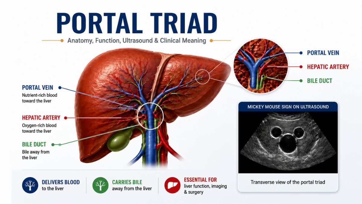
The portal triad is a group of three structures that travel closely together: [1]
- A branch of the portal vein
- A branch of the hepatic artery
- A bile duct
At the entrance to the liver, the larger portal vein, proper hepatic artery, and common bile duct pass through a sheet of tissue called the hepatoduodenal ligament. Inside the liver, progressively smaller branches of these structures travel together within microscopic areas known as portal tracts.
The terms hepatic portal triad and portal triad of the liver refer to the same three-part anatomical arrangement.
However, the portal triad is not the same as the hepatic portal system. The portal triad is a group of three anatomical structures. The hepatic portal system is the wider network of veins that carries blood from the digestive organs and spleen to the liver.
What Does “Portal Triad” Mean in a Medical Report?
In many imaging, surgery, and pathology reports, the term is used simply to identify a normal structure or location.
A report may, for example, state that the portal triad is intact, that blood flow is present, or that inflammation is concentrated around portal tracts. These statements have very different meanings.
Consider asking your healthcare professional:
- Which part of the portal triad is being described?
- Is this normal anatomy, an anatomical variation, or an abnormal finding?
- Is the portal vein open and carrying blood normally?
- Is bile drainage affected?
- Are my liver blood tests abnormal?
- Do I need follow-up imaging or specialist care?
Avoid interpreting one phrase in isolation. Your symptoms, examination, blood-test results, and the rest of the imaging or pathology report all help determine its significance.
What Structures Make Up the Portal Triad?
Each part of the portal triad has a different role. The portal vein and hepatic artery bring blood into the liver, while the bile duct carries bile away.
| Structure | What it carries | Direction | Primary role |
|---|---|---|---|
| Portal vein branch | Nutrient-rich venous blood | Toward the liver | Delivers nutrients, medications, metabolic products, and absorbed substances for processing |
| Hepatic artery branch | Oxygen-rich arterial blood | Toward the liver | Supplies oxygen to liver tissue and the bile ducts |
| Bile duct | Bile produced by liver cells | Away from liver cells | Transports bile toward the gallbladder or small intestine |
Portal vein
The portal vein collects blood from the stomach, intestines, pancreas, spleen, and gallbladder and delivers it to the liver.
This arrangement allows the liver to process substances absorbed from the digestive tract before they reach the wider circulation. These substances may include nutrients, medications, alcohol, toxins, hormones, and metabolic waste.
Approximately 75% of the liver’s blood flow comes through the portal vein. Although this blood contains less oxygen than arterial blood, it is not completely deoxygenated and still contributes to the liver’s oxygen supply.
Hepatic artery
The hepatic artery supplies approximately 25% of the liver’s blood flow.[2] It delivers oxygen-rich blood to liver tissue and is especially important for supporting the bile ducts.
Inside the liver, blood from the hepatic artery and portal vein mixes in tiny, specialized channels called hepatic sinusoids. Liver cells are positioned alongside these channels, giving them close access to oxygen, nutrients, and other substances carried in the blood.
Bile duct
The bile duct carries bile away from the liver.
Bile is a digestive fluid produced by hepatocytes, the liver’s main functional cells. It helps the body digest and absorb fats and provides a pathway for eliminating bilirubin, excess cholesterol, and certain waste products.
Bile initially enters tiny channels called bile canaliculi and then flows into progressively larger bile ducts. The right and left hepatic ducts join to form the common hepatic duct. From there, bile may pass through the cystic duct into the gallbladder for storage or continue through the common bile duct into the duodenum.
Where Is the Portal Triad Located?
The same three-part anatomical arrangement can be described at two scales: the larger structures near the liver’s entrance and their smaller branches within portal tracts inside the liver.
In the hepatoduodenal ligament
The main components of the portal triad travel through the hepatoduodenal ligament, which forms the free edge of the lesser omentum and extends from the porta hepatis toward the upper part of the duodenum.
In the usual anatomical arrangement:
- The portal vein lies toward the back.
- The common bile duct lies toward the front and the patient’s right.
- The proper hepatic artery lies toward the front and the patient’s left.
This compact arrangement is important during liver, gallbladder, pancreatic, and bile duct procedures. However, individual anatomy can vary, which is why surgeons often review detailed imaging before complex operations.
Inside the liver
Under a microscope, the three components of the portal triad lie within connective-tissue areas called portal tracts.[3] The triad itself has three components, while the surrounding portal tract may also contain lymphatic vessels, nerve fibers, and supporting connective tissue.
A portal tract typically contains:
- A portal vein branch, or portal venule
- A hepatic artery branch, or hepatic arteriole
- An interlobular bile duct
- Lymphatic vessels
- Nerve fibers
- Supporting connective tissue
The classic liver lobule is often illustrated as a roughly hexagonal structure. A central vein lies near the middle, while portal tracts sit around the outer edges.
Real liver tissue is less geometrically tidy than a textbook diagram, but the model remains useful for understanding blood flow, bile drainage, and patterns of liver injury.
How Does the Portal Triad Work?
The portal triad provides pathways for two essential processes: delivering blood toward the liver sinusoids and draining bile away from liver cells.
Blood from the portal vein and hepatic artery enters the hepatic sinusoids. As the blood passes liver cells, the liver can:
- Process carbohydrates, fats, and proteins
- Store glucose, vitamins, and minerals
- Metabolize medications and alcohol
- Modify or remove potentially harmful substances
- Produce albumin, clotting factors, and other proteins
- Regulate several hormones and metabolic processes
After passing through the sinusoids, the blood drains into central veins, hepatic veins, and finally the inferior vena cava, which returns it to the general circulation.
The simplified blood-flow pathway is:
Portal vein and hepatic artery branches → sinusoids → central veins → hepatic veins
Bile generally travels in the opposite direction:
Hepatocytes → bile canaliculi → small bile ducts → larger hepatic ducts
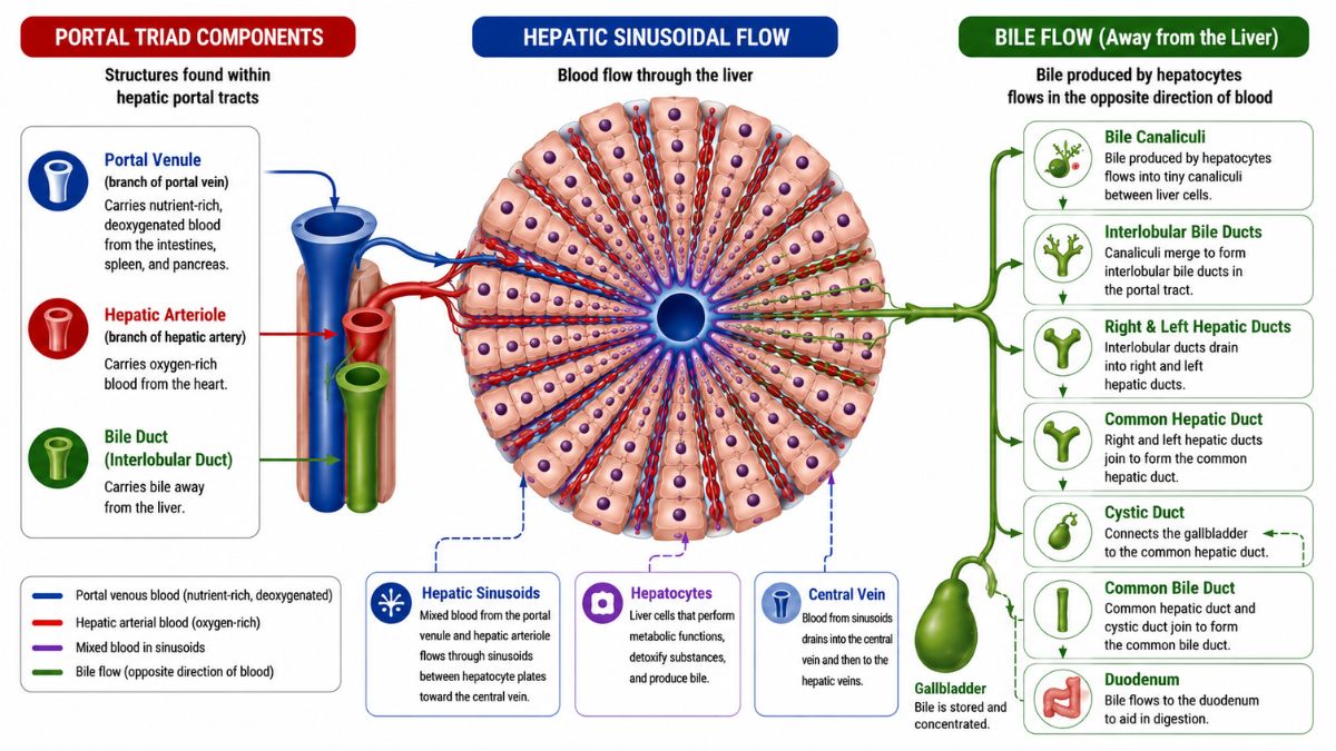
At the microscopic level, bile flows toward the portal tracts, opposite to the direction of blood flowing toward the central veins. This opposing movement is often described as countercurrent flow.
What Does the Portal Triad Look Like on Ultrasound?
On a transverse ultrasound image near the liver’s entrance, the portal triad may create a recognizable appearance called the Mickey Mouse sign.[4]
In this view:
- The portal vein forms the larger posterior “head.”
- The hepatic artery forms one smaller anterior “ear.”
- The common bile duct forms the other anterior “ear.”
This sign is an anatomical landmark—not an abnormal finding. The structures may not always form a perfect shape because ultrasound appearance depends on the scanning angle and individual anatomy.
Color Doppler ultrasound can help distinguish the three structures. With appropriately optimized Doppler settings, blood flow is typically detectable in the portal vein and hepatic artery, while the bile duct normally shows no internal vascular flow.
Depending on the clinical question, imaging may help evaluate:
- Portal vein thrombosis, or a blood clot in the portal vein
- Abnormal portal blood flow
- Bile duct widening or obstruction
- Masses around the porta hepatis
- Changes related to portal hypertension
CT, particularly CT angiography when detailed vascular mapping is needed, can provide clearer views of portal and arterial anatomy. MRI and magnetic resonance cholangiopancreatography, commonly called MRCP, are especially useful for evaluating the bile ducts and surrounding soft tissues.[5]
Why Is the Portal Triad Clinically Important?
The portal triad is important in liver surgery, transplantation, trauma care, diagnostic imaging, and liver-biopsy interpretation because disease or injury affecting one component can alter blood flow, oxygen delivery, or bile drainage.
Anatomical variations
Not everyone has the same portal vein branching pattern—and variation does not automatically mean something is wrong.
A 2024 systematic review and meta-analysis included 21 studies and 51,244 subjects.[6] The pooled estimates suggested that:
- Portal vein trifurcation occurred in about 8%
- A right posterior portal vein arising directly from the main portal vein occurred in about 7%
- A right anterior portal vein arising from the left portal vein occurred in about 4%
- Other isolated variants occurred in about 2%
The authors reported substantial variation between the included studies, so these pooled percentages should be interpreted as broad estimates rather than fixed rates that apply equally to every population.
Most anatomical variants do not cause symptoms. They become especially important when a procedure depends on the exact position of a vessel or bile duct, such as:
- Liver resection
- Liver transplantation
- Portal vein embolization
- Tumor ablation
- Complex bile duct surgery
The Pringle maneuver
During major liver bleeding or certain liver operations, a surgeon may temporarily compress the hepatoduodenal ligament. This technique is called the Pringle maneuver.[7]
Because the ligament contains the portal vein and hepatic artery, compression reduces blood flowing into the liver and may help control bleeding.
The maneuver does not stop bleeding from the hepatic veins or inferior vena cava, which are part of the liver’s outflow rather than the portal triad.
Conditions involving portal-triad structures
Medical reports may discuss the portal triad or portal tracts when evaluating:
- Portal vein thrombosis
- Portal hypertension
- Bile duct obstruction
- Cholangitis, or bile duct inflammation or infection
- Periportal inflammation
- Periportal fibrosis
- Liver or bile duct tumors
- Masses near the porta hepatis
- Changes following liver, gallbladder, or bile duct surgery
Terms such as patent or normal flow are usually reassuring. Words such as dilated, obstructed, thrombosed, inflamed, fibrotic, or infiltrated describe findings that may require further interpretation.
Advanced Anatomy: Portal Triad Histology and Liver Zones
The portal triad also helps explain the hepatic acinus, a model that divides liver tissue into three functional zones.
These zones receive different amounts of oxygen and have different metabolic roles. As a result, certain liver injuries affect one zone more than another.
Zone 1: Periportal zone
Zone 1 lies closest to the portal tract and receives incoming blood first. It generally has the highest oxygen supply.
Cells in this zone are active in oxygen-dependent processes and are among the first to encounter nutrients, medications, toxins, and gut-derived microbial products carried in portal blood. However, the location of drug-induced liver injury depends on how a substance is metabolized and is not always greatest in Zone 1.
Zone 2: Intermediate zone
Zone 2 sits between Zones 1 and 3. Its oxygen supply, metabolic activity, and vulnerability to injury are generally intermediate.
Zone 3: Centrilobular zone
Zone 3 is closest to the central vein and receives blood after it has passed through the other zones. It therefore has the lowest relative oxygen supply.
This zone contains substantial activity from the cytochrome P450 enzyme system, which helps metabolize many medications and chemicals. Consequently, Zone 3 may be particularly vulnerable to reduced blood flow and to substances that become toxic after metabolism.
Doctors and pathologists use these patterns to interpret liver biopsy findings and understand how different conditions affect liver tissue.
When Liver or Bile-Duct Symptoms Need Urgent Care
The words “portal triad” are not themselves an emergency. However, conditions affecting liver blood flow, bile drainage, or nearby structures can sometimes cause symptoms that require urgent assessment.
Seek prompt emergency care for:
- Yellowing of the skin or eyes together with fever, chills, or right-upper-abdominal pain
- Vomiting blood
- Black, tarry, or visibly bloody stools
- New confusion, unusual drowsiness, or loss of consciousness
- Fainting, severe weakness, or signs of shock
- Severe or rapidly worsening abdominal pain
Fever, worsening jaundice, and pain in the upper-right abdomen can occur with a serious bile duct infection.[8] Vomiting blood or passing black stools may indicate significant gastrointestinal bleeding.
These symptoms do not prove that there is a portal-triad disorder, but they should not be assessed through online information alone.
Frequently Asked Questions
The portal triad itself contains three structures: a portal vein branch, a hepatic artery branch, and a bile duct. The surrounding portal tract may also contain lymphatic vessels, nerves, and connective tissue.
It provides routes for portal venous and arterial blood to enter the liver while allowing bile to drain away from liver cells.
No. It is normal anatomy. A disease may affect one or more of its components, but the term itself is not a diagnosis.
No. The porta hepatis is the liver’s gateway or hilum. The main structures of the portal triad pass through this area.
No. The portal triad is a group of three structures. The hepatic portal system is the network of veins transporting blood from the digestive organs and spleen to the liver.
Yes. In a transverse view, it may form the Mickey Mouse sign, with the portal vein appearing as the head and the hepatic artery and bile duct appearing as the ears.
Usually not. Many variations are present from birth and cause no symptoms. They mainly matter when planning liver, vascular, transplant, or bile duct procedures.
The Bottom Line
The portal triad is a normal anatomical arrangement consisting of a portal vein branch, a hepatic artery branch, and a bile duct. Together, these structures deliver blood to the liver and carry bile away.
Seeing the term “portal triad” in a medical report does not automatically mean that something is wrong. Its significance depends on the surrounding findings and whether blood flow, bile drainage, or nearby tissues are affected.
Medical References
Update History
Medically reviewed by Dr. Rahul Kushwaha.
Originally written by Dr. Jubair Islam.
Your answer helps us improve the clarity and usefulness of our health content.