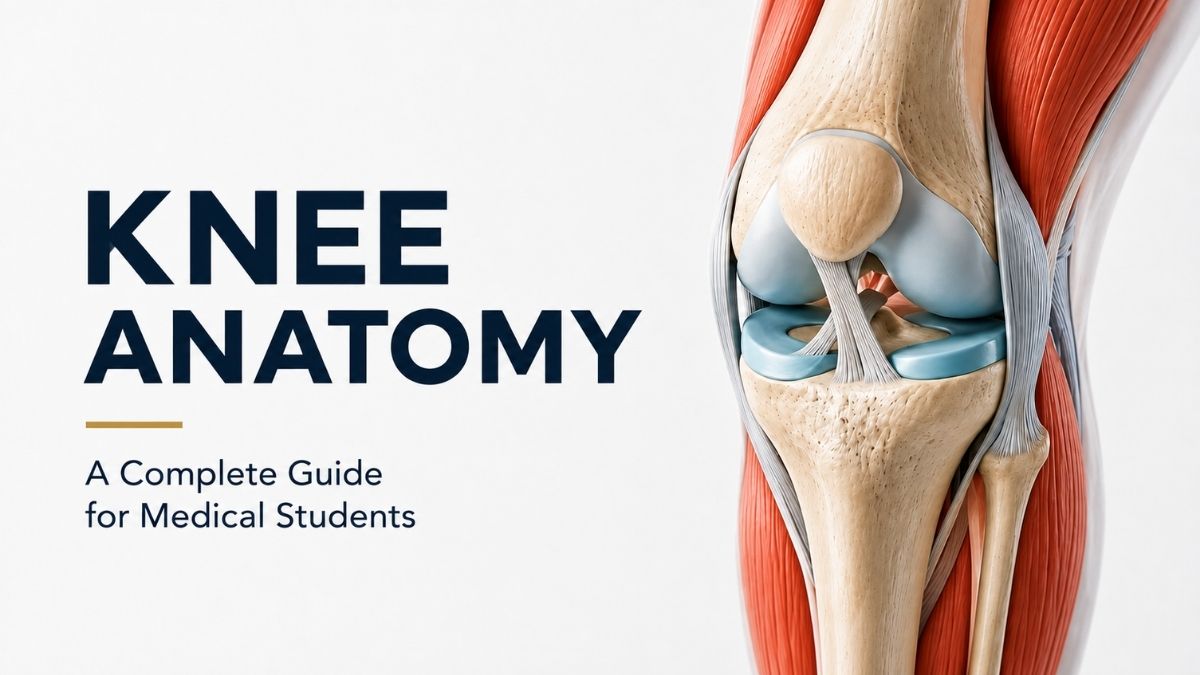
The knee looks like a hinge—until you watch it move.
During a squat, sprint, or even a simple step, the joint does far more than flex and extend. The femur rolls and glides over the tibia, the menisci deform under load, the patella shifts its contact with the trochlea, and ligaments tighten or relax as joint position changes. Muscles add another layer of active control.
That complexity is also why a small change in one structure can alter the mechanics of the entire joint.
This is the most useful way to learn knee anatomy: not as a list of structures to memorize, but as a coordinated mechanical system in which anatomy, movement, stability, and injury are closely connected.
The knee is generally considered the largest synovial joint in the body and is commonly described as a modified hinge or hinge-type synovial joint. Its major articulations are the tibiofemoral and patellofemoral joints. Flexion and extension dominate its movement, but axial rotation, translation, rolling, and gliding are essential to normal function.[1]
For medical students, the goal is bigger than memorizing what attaches where. The more useful question is: What does each structure control, and what changes when it is injured?
Knee Anatomy at a Glance
| Structure | Key Anatomy | Main Functional Role |
|---|---|---|
| Femur | Medial and lateral condyles, trochlea | Forms the superior tibiofemoral surfaces and patellofemoral groove |
| Tibia | Medial and lateral plateau, intercondylar region, tibial tuberosity | Supports the femur and transmits load |
| Patella | Sesamoid bone within the extensor mechanism | Increases the quadriceps moment arm and guides extensor forces |
| Articular cartilage | Hyaline cartilage covering joint surfaces | Provides a smooth, low-friction, load-bearing surface |
| Menisci | Medial and lateral fibrocartilage | Improve congruence, distribute load, and contribute to stability |
| ACL and PCL | Intracapsular, extrasynovial cruciate ligaments | Control anterior-posterior translation and contribute to rotational stability |
| MCL and LCL | Major collateral stabilizers | Resist excessive valgus and varus movement |
| Muscles and tendons | Quadriceps, hamstrings, popliteus, gastrocnemius, and others | Produce movement and provide dynamic stability |
| Capsule and synovium | Fibrous capsule lined by synovium | Enclose, reinforce, and lubricate the joint |
| Neurovascular structures | Genicular vessels and articular nerve branches | Supply tissues, sensation, proprioceptive input, and motor control |
Bones and Articulations of the Knee
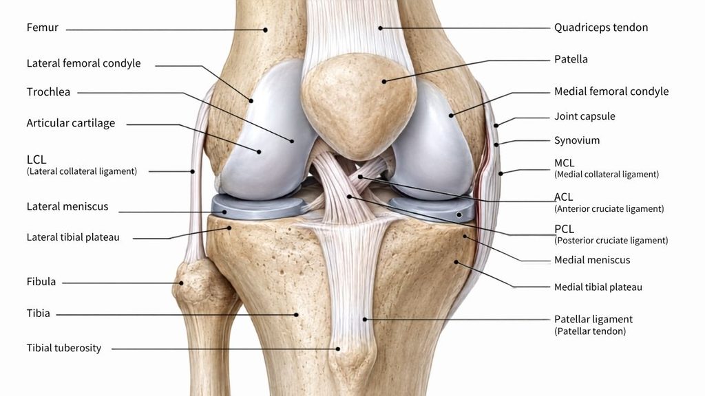
Three bones form the principal knee joint: the femur, tibia, and patella.
The fibula is closely related to the knee but does not articulate directly with the femur or form part of the main tibiofemoral articulation. Its head forms the proximal tibiofibular joint with the tibia and provides important attachment sites for the fibular collateral ligament (LCL) and biceps femoris tendon.
Femur
The distal femur expands into the medial and lateral femoral condyles. These curved surfaces articulate with the tibial plateau and are covered by hyaline articular cartilage.
Between the condyles posteriorly is the intercondylar fossa, closely related to the cruciate ligaments. Anteriorly, the condyles blend into the femoral trochlea, where the patella tracks during flexion and extension.
The two condyles are not geometrically identical. That asymmetry matters because it contributes to the coupled rotation seen near terminal knee extension.
Tibia
The proximal tibia forms the medial and lateral tibial plateaus. Compared with the curved femoral condyles, these surfaces are relatively flat, so the knee has limited inherent bony congruence.
This limited bony congruence is partly compensated for by the menisci.
Between the plateaus lies the intercondylar region, which contains attachment areas for the cruciate ligaments and meniscal roots. Anteriorly, the tibial tuberosity receives the patellar ligament and marks the distal end of the extensor mechanism.
Patella
The patella is the largest sesamoid bone in the human body. It lies within the quadriceps extensor mechanism and articulates posteriorly with the femoral trochlea.
Beyond protecting the anterior knee, the patella holds the extensor mechanism farther from the knee’s axis of rotation, increasing the quadriceps moment arm and improving the mechanical efficiency of knee extension.
Tibiofemoral Joint
The tibiofemoral articulation can be considered in medial and lateral compartments. Each contains a femoral condyle, the corresponding tibial plateau, and a meniscus.
The medial side is relatively constrained, while the lateral compartment allows greater mobility. This asymmetry becomes especially important during flexion and rotation.
Patellofemoral Joint
The patellofemoral joint lies between the posterior surface of the patella and the femoral trochlea.
Patellofemoral contact is not fixed. As knee flexion changes, different portions of the patella contact the femur.
Normal tracking depends on several factors working together, including trochlear geometry, patellar shape, limb alignment, retinacular tissues, and the quadriceps-extensor mechanism. The medial patellofemoral ligament (MPFL) is an important passive restraint to lateral displacement of the patella, particularly in early knee flexion.
Articular Cartilage and Menisci
The word cartilage can be misleading around the knee because two structurally different tissues are involved.
Articular cartilage is predominantly hyaline cartilage, whereas the menisci are fibrocartilaginous structures. Their composition and mechanical functions are therefore not interchangeable.
Articular Cartilage
Articular cartilage covers the femoral condyles, tibial articular surfaces, and posterior surface of the patella.
It is highly specialized for repeated loading while allowing remarkably low-friction movement.
Histologically, mature articular cartilage is commonly divided into superficial, transitional, deep or radial, and calcified zones. Collagen orientation, chondrocyte arrangement, water content, and proteoglycan concentration vary by depth.
This zonal architecture allows the tissue to tolerate both shear and compressive forces.
Articular cartilage is also avascular and aneural. Nutrient movement therefore depends largely on diffusion, and its intrinsic ability to repair substantial defects is limited.
Medial vs. Lateral Meniscus
The medial meniscus is generally more C-shaped and more firmly attached to surrounding capsular structures. Its relationship with the deep medial collateral ligament contributes to its relative lack of mobility.
The lateral meniscus is more nearly circular and generally more mobile. Its capsular attachment is interrupted around the popliteal hiatus, and it does not have the same broad attachment relationship with the LCL that the medial meniscus has with medial structures.
This difference in mobility is functionally important because the menisci must adapt as femorotibial contact changes throughout knee motion.
What Do the Menisci Actually Do?
Calling the menisci “shock absorbers” is not wrong, but it is incomplete.
They:
- Increase joint congruence
- Distribute contact loads
- Contribute to stability
- Assist joint lubrication
- Participate in proprioception
- Help transmit compressive forces across the tibiofemoral joint
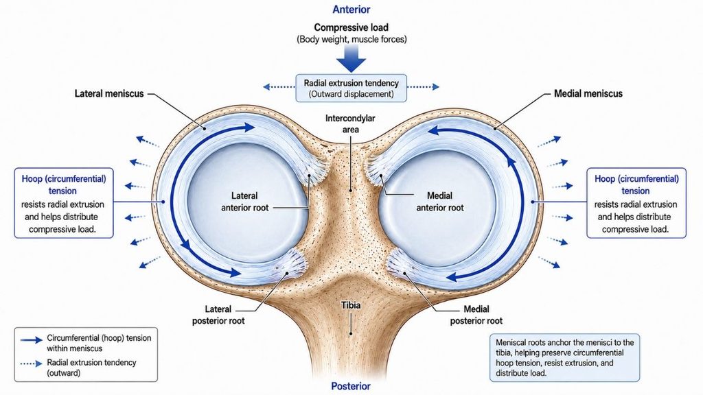
Their wedge shape and collagen architecture allow compressive loads to be converted into circumferential tensile forces. Circumferential collagen fibers are especially important for resisting this outward expansion, while radial fibers help stabilize the collagen network and resist longitudinal splitting.[2]
This circumferential tension is commonly called hoop stress or hoop tension.
Meniscal Roots and Hoop Stress
Each meniscus is anchored to the tibia by anterior and posterior roots.
These attachments prevent excessive outward displacement, or extrusion, when the joint is loaded.
If an important meniscal root fails, normal hoop-stress transmission is compromised. Contact mechanics across the tibiofemoral joint can then change substantially.
That is why a relatively small root tear can have a disproportionately large biomechanical effect.
A useful principle to remember is:
The meniscus can function as an effective load-distribution system only when its structure and attachments remain mechanically connected.
Meniscal Vascular Zones
Blood supply is greatest in the peripheral portion of the adult meniscus and decreases toward the inner free edge.
For teaching purposes, vascularity is traditionally described in three zones:
- Red-red zone: vascular peripheral region
- Red-white zone: transitional region
- White-white zone: relatively avascular inner region
These zones are not perfectly sharp anatomical boundaries. Vascular penetration also varies with age and between individuals.
Clinically, the distinction matters because vascular supply influences healing potential following meniscal injury.
Ligaments and Stabilizing Complexes
The ACL, PCL, MCL, and LCL are the four ligaments most medical students learn first.
That is a useful starting point—but it is not the whole stability system.
Ligament function is also position-dependent. A major knee ligament rarely controls only one type of motion.
Anterior Cruciate Ligament
The anterior cruciate ligament (ACL) arises from the anterior intercondylar region of the tibia and runs superiorly, posteriorly, and laterally toward the lateral femoral condyle.
Its best-known role is limiting excessive anterior translation of the tibia relative to the femur, but the ACL also contributes strongly to rotational stability.
That second role helps explain why an ACL-deficient knee may feel unstable during cutting or pivoting movements even when anterior translation alone does not explain the patient’s symptoms.
Posterior Cruciate Ligament
The posterior cruciate ligament (PCL) runs from the posterior intercondylar region of the tibia toward the medial femoral condyle.
Its major role is resisting excessive posterior translation of the tibia relative to the femur, particularly when the knee is flexed, while also contributing to rotational control.
Both cruciate ligaments lie within the fibrous capsule, while the synovial membrane excludes them from the main synovial cavity. Anatomists therefore classify them as intracapsular but extrasynovial.
Medial Collateral Ligament
The medial collateral ligament is better understood as a complex than as a single flat band.
Its superficial component is the major restraint to excessive valgus loading.
Deeper medial structures are closely related to the joint capsule and medial meniscus and also contribute to rotational stability.
This layered anatomy helps explain why significant medial-sided injuries may involve more than one anatomical structure.
Lateral Collateral Ligament
The lateral collateral ligament, also called the fibular collateral ligament, extends from the lateral femoral region to the fibular head.
Its major role is resisting excessive varus stress.
Unlike the medial collateral complex, the LCL does not broadly blend with the lateral meniscus.
The Anterolateral Complex
Research on the anterolateral side of the knee shows how anatomical descriptions and understanding can evolve as evidence improves.
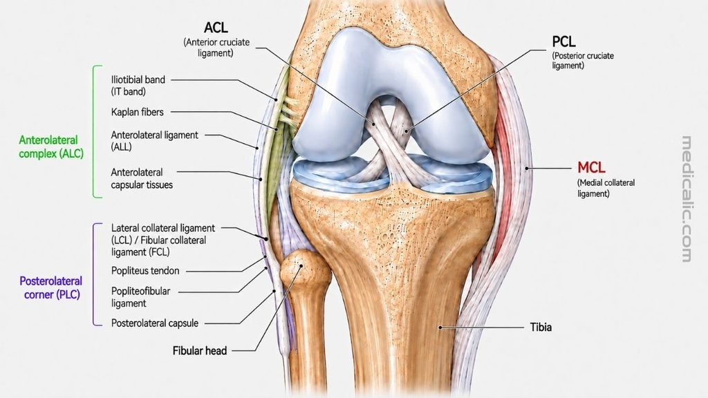
The anterolateral complex (ALC) includes several structures that contribute to rotational control, including the iliotibial tract, its deeper distal attachments such as Kaplan fibers, anterolateral capsular tissues, and the anterolateral ligament.
For examinations, learn the individual structures; for clinical reasoning, remember the broader principle: anterolateral rotational stability is produced by a network, not by one isolated ligament.[3]
Posterolateral Corner
The posterolateral corner is another functionally important stabilizing complex.
Key structures include:
- Fibular collateral ligament
- Popliteus tendon
- Popliteofibular ligament
- Posterolateral capsular structures
Together, they help resist varus loading, excessive external rotation, and posterolateral instability.
This is why injury to relatively small posterolateral structures can have a major effect on knee mechanics.[4]
Muscles, Tendons, and the Extensor Mechanism
Ligaments provide passive restraint, while muscles add dynamic stability by continuously adjusting force as the knee moves and bears load.
Quadriceps and Knee Extension
The quadriceps femoris consists of:
- Rectus femoris
- Vastus medialis
- Vastus lateralis
- Vastus intermedius
Their fibers converge into the quadriceps tendon and transmit force to the patella.
From the inferior pole of the patella, force continues to the tibial tuberosity through the patellar ligament.
Clinically, the term patellar tendon is widely used, while anatomical terminology commonly favors patellar ligament. The structure forms the distal continuation of the quadriceps extensor mechanism from the patella to the tibial tuberosity.
Together, the quadriceps tendon, patella, patellar ligament, and adjacent retinacular tissues form the extensor mechanism.
Hamstrings
The hamstrings are major knee flexors and also influence rotation when the knee is flexed.
Semitendinosus and semimembranosus contribute to medial rotation of the tibia.
Biceps femoris contributes to lateral rotation.
Because several hamstring muscles cross both the hip and knee, their mechanical effects change with limb position.
Popliteus
The popliteus is a small but functionally important muscle of the posterior knee.
It contributes to flexion and rotational control and helps initiate movement away from terminal knee extension.
This is the classic “unlocking” function.
In an open-chain knee, the popliteus can internally rotate the tibia relative to the femur.
In a closed-chain position, it can assist external rotation of the femur relative to the fixed tibia.
Gastrocnemius
The gastrocnemius crosses the posterior knee before continuing distally toward the Achilles tendon.
Although it is best known for plantar flexion at the ankle, it can also assist knee flexion and influence posterior knee mechanics.
Pes Anserinus
The pes anserinus is formed by the tendons of:
- Sartorius
- Gracilis
- Semitendinosus
These tendons insert on the anteromedial proximal tibia.
A useful mnemonic is SGT: sartorius, gracilis, semitendinosus.
Iliotibial Band
The iliotibial band is a thick fascial structure running along the lateral thigh.
Distally, the iliotibial band inserts primarily at Gerdy’s tubercle on the anterolateral proximal tibia and also has important deeper attachments around the distal femur, including Kaplan fiber attachments.
These superficial and deep connections contribute to lateral and anterolateral rotational stability.
Joint Capsule, Synovium, Bursae, and Fat Pads
The knee’s fibrous capsule is not a perfectly uniform sleeve.
Instead, it blends with and is reinforced by surrounding ligaments, tendons, and fascial structures.
Its inner surface is lined by synovium, which produces synovial fluid and helps maintain the nutritional environment of avascular articular cartilage.
Suprapatellar Recess
Superiorly, the synovial cavity extends above the patella as the suprapatellar recess.
This space accommodates movement of the extensor mechanism during knee flexion and extension.
Major Bursae
Numerous bursae reduce friction where tendons, ligaments, skin, and bone move relative to one another.
Clinically important examples include:
- Prepatellar bursa
- Superficial infrapatellar bursa
- Deep infrapatellar bursa
- Pes anserine bursa
- Gastrocnemius-semimembranosus bursal region
Not all bursae communicate with the main knee joint cavity.
Hoffa’s Fat Pad
The infrapatellar fat pad, commonly called Hoffa’s fat pad, lies deep to the patellar ligament.
It is intracapsular but extrasynovial and occupies changing spaces as the knee moves.
Because it has a rich neurovascular supply, it may also become an important source of anterior knee pain.
Blood Supply and Innervation
The posterior knee is more than a collection of muscles and ligaments; it is also a major neurovascular corridor.
Arterial Supply
The knee receives blood through an extensive periarticular network commonly referred to as the genicular anastomosis.
Important arterial contributors arise from the popliteal system, with additional input from femoral and recurrent vessels.
This periarticular network supplies several knee structures, including:
- Capsule
- Synovium
- Cruciate ligaments
- Peripheral meniscal tissue
- Bone
- Surrounding soft tissue
The popliteal artery lies deep within the popliteal fossa directly behind the knee, making its anatomical relationship particularly important during major trauma and surgery. In the central popliteal fossa, the major neurovascular structures are arranged roughly from superficial to deep as the tibial nerve, popliteal vein, and popliteal artery.
Nerve Supply
Sensory innervation of the knee is more complex than diagrams showing only a handful of “genicular nerves” may suggest.
Articular branches arise from multiple pathways associated with the femoral, saphenous, tibial, common fibular, and obturator nerves.
The common fibular nerve also has an important superficial relationship to the knee: it passes around the neck of the fibula, where it is particularly vulnerable to compression or injury.[5]
There is considerable anatomical variation in articular innervation.
For medical students, one distinction is especially useful:
Motor innervation of muscles and sensory innervation of the joint are related, but they are not identical systems.
A nerve supplying a muscle that moves the knee may also contribute articular branches to the joint. This relationship reflects Hilton’s law, which broadly states that nerves supplying muscles acting on a joint often also provide sensory branches to that joint.
How the Knee Moves: Functional Biomechanics
Unlike a simple hinge, the knee does not move around one fixed axis. Flexion and extension dominate, but normal motion also includes axial rotation, anterior-posterior translation, small varus-valgus movements, rolling, and gliding.[6]
Flexion and Extension
As the knee flexes, contact between the femoral condyles, tibial surfaces, and menisci continuously changes.
The menisci move with the changing contact surfaces, and ligament tension varies throughout the range of motion.
As the knee approaches full extension, the joint becomes progressively more stable.
The Screw-Home Mechanism
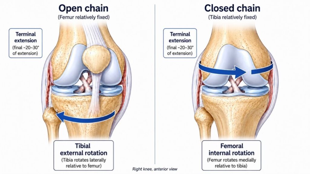
Near terminal extension, a small amount of coupled rotation occurs.
In an open-chain movement, the tibia externally rotates relative to the femur as the knee reaches full extension.
In a weight-bearing closed-chain position, the equivalent motion is internal rotation of the femur relative to the tibia.
This is called the screw-home mechanism.
Several factors contribute, including:
- Femoral condylar geometry
- Ligament tension
- Articular surface shape
- Muscle activity
The popliteus helps reverse this rotational relationship when flexion begins.
The knee does not literally “lock” like a mechanical bolt. Rather, terminal extension creates a more stable configuration that requires coordinated rotation as flexion begins.
Roll and Glide
The femoral condyles cannot simply roll across the tibial plateau; pure rolling would quickly carry them beyond the available articular surface. Instead, rolling and gliding occur together, with their relative contribution changing across the range of motion and depending partly on whether the femur or tibia is moving. This combination preserves articular contact while allowing substantial flexion.
Meniscal Movement
The menisci deform and translate as tibiofemoral contact changes through motion. The lateral meniscus is generally more mobile than the medial because of differences in capsular attachment and its relationship with the popliteus, helping it accommodate changing joint contact while maintaining load distribution.
Joint loading also changes with lower-limb alignment. Varus alignment tends to shift relatively more load toward the medial tibiofemoral compartment, whereas valgus alignment shifts relatively more load laterally. The effect in an individual knee also depends on muscle forces, meniscal position, and overall joint geometry.
Knee Anatomy on X-Ray and MRI
Dissection teaches anatomical relationships; imaging teaches you to recognize those same relationships in a living patient.
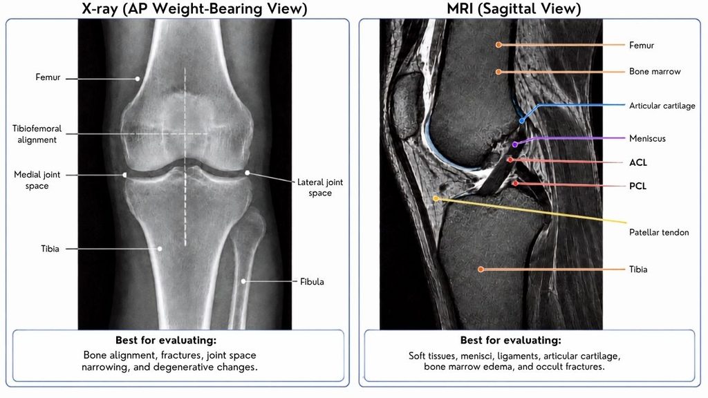
X-Ray
Plain radiography is particularly useful for evaluating:
- Bone
- Alignment
- Fracture
- Osteophytes
- Joint-space relationships
- Patellofemoral alignment
Articular cartilage and menisci are not directly visible on standard X-rays.
Changes in joint-space width are therefore interpreted indirectly in the context of cartilage, meniscal, and bony changes.
Weight-bearing frontal views are useful for assessing tibiofemoral compartments and alignment. Lateral and axial or skyline views provide additional information about the patellofemoral joint.
MRI
MRI provides far greater soft-tissue detail.
It can visualize structures including:
- Menisci
- ACL and PCL
- Collateral ligaments
- Articular cartilage
- Tendons
- Bone marrow
- Muscles
- Capsule
- Fat pads
For students, thinking in the three standard MRI planes makes anatomy much easier.
Sagittal images are particularly useful for the cruciate ligaments, extensor mechanism, and meniscal horns.
Coronal images help define the collateral ligaments, meniscal bodies, and medial-lateral compartment anatomy.
Axial images are especially useful for patellofemoral relationships and transverse soft-tissue anatomy.[7]
A practical study technique is to follow the same structure through all three planes rather than memorizing each plane separately.
Anatomy becomes three-dimensional much faster that way.
Normal Anatomical Variation and Advanced Imaging
Textbook illustrations are useful models, but normal human knees do not follow a single anatomical blueprint.
Variation can occur in:
- Meniscal attachments
- Meniscofemoral ligaments
- Patellar morphology
- Lateral meniscal structures
- Fabella presence
- Accessory ligamentous structures
The meniscofemoral ligaments connect the posterior horn of the lateral meniscus with the femur in close relationship to the PCL. When present, the anterior meniscofemoral ligament passes anterior to the PCL, and anatomists commonly call it the ligament of Humphrey. The posterior meniscofemoral ligament passes posterior to the PCL and is known as the ligament of Wrisberg.
Another important variant is the discoid meniscus, a broader, more disc-like meniscal morphology that most commonly affects the lateral meniscus. It may be asymptomatic or associated with mechanical symptoms.
Ultra-high-field imaging has reinforced an important anatomical lesson: normal knees show meaningful structural variation. In one high-resolution 7-tesla MRI study of 57 asymptomatic knees, the posterior root of the lateral meniscus showed variable insertion patterns. These findings remind students that textbook anatomy is a model rather than an exact blueprint. Seven-tesla MRI remains largely research-oriented, while conventional MRI continues to be the clinical workhorse for routine knee imaging.[8]
Why Knee Anatomy Matters Clinically
The easiest way to turn knee anatomy into clinical reasoning is to ask what movement or stabilizing function changes when a structure fails.
ACL Injury
Because the ACL restrains both anterior translation and rotational motion, injury may produce instability that becomes especially apparent during pivoting or cutting movements.
Meniscal Injury
Meniscal injury can disrupt normal load distribution, with root tears being particularly important because loss of tibial attachment can promote extrusion and markedly alter joint mechanics.
MCL and LCL Injury
The MCL is the major restraint to excessive valgus loading.
The LCL and surrounding posterolateral structures contribute substantially to resistance against varus and rotational instability.
Posterolateral Corner Injury
Because the posterolateral corner contributes to varus, rotational, and posterolateral stability, injury can disrupt several directions of control at once.
Patellofemoral Problems
Patellar tracking depends on the interaction of patellar morphology, trochlear geometry, limb alignment, the extensor mechanism, retinacular tissues, and surrounding ligaments.
Patellofemoral disorders therefore rarely result from one structure acting alone. Injury to medial stabilizers such as the MPFL is particularly relevant after lateral patellar dislocation, while trochlear morphology, alignment, and extensor mechanics can influence recurrent instability.
Osteoarthritis
Knee osteoarthritis is more than “worn cartilage.” It can involve articular cartilage, menisci, subchondral bone, synovium, ligaments, alignment, and the overall joint architecture, making it better understood as a disease of the entire joint.[9]
Once these structure-function relationships become clear, knee anatomy becomes less about memorization and more like a clinical language for understanding examination findings, imaging, and injury patterns.
Frequently Asked Questions About Knee Anatomy
The major structures include the femur, tibia, patella, articular cartilage, medial and lateral menisci, ACL, PCL, MCL, LCL, capsule, synovium, muscles, tendons, bursae, blood vessels, and articular nerves.
Together, they provide mobility, stability, load transmission, and sensory feedback.
The principal articulating bones are the femur, tibia, and patella.
The fibula is closely related to the knee and provides important ligament and tendon attachment sites, but it does not articulate directly with the femur.
The four major ligaments traditionally emphasized are the ACL, PCL, MCL, and LCL. The cruciate ligaments are particularly important for anterior-posterior and rotational stability, while the collateral ligaments primarily resist excessive valgus and varus loading.
The medial meniscus is more C-shaped, firmly attached, and relatively less mobile. The lateral meniscus is more circular and mobile, partly because its capsular attachment is interrupted by the popliteal hiatus.
Yes, but only partly. The knee is a modified hinge synovial joint: flexion and extension dominate, while rotation, translation, rolling, and gliding are also essential to normal movement.
Key Takeaways
Understanding knee anatomy becomes easier when each structure is linked to its function. Bones provide the framework, cartilage and menisci manage joint contact and load, ligaments guide stability, and muscles add dynamic control.
Most importantly, the knee is not a static hinge. It is a three-dimensional mechanical system in which joint shape, tissue tension, muscle force, and position continuously influence one another.
Learn those relationships, and anatomy, imaging, physical examination, and injury patterns begin to connect into the same clinical picture.
Medical References
Update History
Originally written by Dr. Jubair Islam.
Your answer helps us improve the clarity and usefulness of our health content.